This Village-Made Chinese Pain Reliever Eliminates Back And Joint Pain!
Exploring Temporomandibular Joint Anatomy: What You Need to Know

Introduction to the Temporomandibular Joint
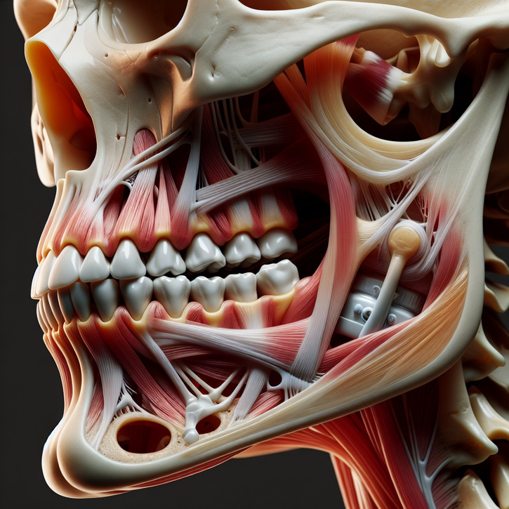
The temporomandibular joint, often abbreviated as TMJ, plays a crucial role in our daily lives, yet it is frequently overlooked until problems arise. This complex joint connects the jawbone to the skull, enabling essential functions such as speaking and chewing. Understanding the intricacies of TMJ anatomy is vital for both medical professionals and individuals experiencing discomfort in this area. TMJ disorders can affect quality of life, making it important to be informed about its structure and function. This guide delves into the anatomy of the temporomandibular joint, exploring its components, functions, and related muscles.
Overview of the Temporomandibular Joint
The temporomandibular joint is a sophisticated hinge that allows the jaw to move smoothly. It is located on each side of the head, just in front of the ears. This joint is unique as it combines a hinge action with sliding motions, making it one of the most complex joints in the body. The TMJ facilitates a wide range of jaw movements, including opening, closing, and side-to-side motion. Its complexity is due to the intricate interplay of bones, muscles, ligaments, and nerves that work in harmony to ensure efficient jaw function.
Importance of Understanding TMJ Anatomy
Understanding the anatomy of the temporomandibular joint is crucial for diagnosing and treating TMJ disorders. Healthcare professionals, such as dentists and otolaryngologists, rely on this knowledge to identify the root causes of TMJ pain and dysfunction. For individuals experiencing jaw pain or other related symptoms, being informed about TMJ anatomy can aid in seeking appropriate treatment. Moreover, a clear understanding of TMJ anatomy can assist in preventing potential issues by promoting healthy habits and early intervention. This knowledge empowers individuals to take proactive steps towards maintaining optimal jaw health.
Basic Anatomy of the Temporomandibular Joint
Components of the Temporomandibular Joint
The temporomandibular joint is composed of several key components that work together to facilitate jaw movement. These include the mandibular condyle, the articular disc, the temporal bone, and the joint capsule. The mandibular condyle is the rounded end of the lower jawbone that fits into the temporal bone's socket. Between these two bones lies the articular disc, a flexible cartilage that cushions and stabilizes the joint during movement. The joint capsule encloses the TMJ, providing support and protection. Each component plays a vital role in ensuring smooth and coordinated jaw function.
Functions of the TMJ
The temporomandibular joint serves several essential functions in our daily lives. Primarily, it allows for the opening and closing of the mouth, which is crucial for speaking, eating, and expressing emotions. Additionally, the TMJ enables the jaw to move side-to-side and forward and backward, allowing for a wide range of oral activities. This joint's ability to perform these complex movements is attributed to its unique structure and the coordination of surrounding muscles and ligaments. The TMJ's intricate functionality is fundamental to maintaining proper oral health and overall wellbeing.
Role of TMJ in Jaw Movement
The temporomandibular joint is pivotal in facilitating various jaw movements essential for daily activities. It allows for the hinge movement necessary for opening and closing the mouth, as well as the gliding motion required for chewing and speaking. The TMJ's unique design enables these movements to occur smoothly and efficiently. This joint's flexibility is crucial for performing tasks such as biting, grinding, and yawning without discomfort. Any disruption in the TMJ's function can lead to difficulties in jaw movement, highlighting the importance of its proper maintenance and care.
Muscles Involved in Temporomandibular Joint Function
Primary Muscles Supporting TMJ
The primary muscles supporting the temporomandibular joint are the masseter, temporalis, medial pterygoid, and lateral pterygoid muscles. These muscles are responsible for the major movements of the jaw, such as opening, closing, and side-to-side motion. The masseter and temporalis muscles are powerful and primarily involved in closing the jaw, while the medial and lateral pterygoid muscles assist in opening the jaw and facilitating complex movements. The coordinated effort of these muscles ensures smooth and efficient jaw function, highlighting their critical role in maintaining TMJ health.
Secondary Muscles Affecting TMJ
In addition to the primary muscles, several secondary muscles influence the function of the temporomandibular joint. These include the digastric, mylohyoid, and geniohyoid muscles, which assist in opening the jaw and stabilizing the hyoid bone during movement. The sternocleidomastoid and trapezius muscles also play a role in supporting the TMJ by maintaining proper head and neck posture. Although these muscles are not directly involved in jaw movement, their coordination and balance are essential for optimal TMJ function and preventing disorders related to muscle tension or imbalance.
Muscle Coordination in TMJ Movement
Muscle coordination is vital for the proper functioning of the temporomandibular joint. The harmonious interaction between the primary and secondary muscles ensures smooth and controlled jaw movements. This coordination is regulated by the central nervous system, which sends signals to the muscles, enabling them to contract and relax as needed. Proper muscle coordination prevents excessive strain on the TMJ and reduces the risk of disorders. Regular exercises and stretches can enhance muscle coordination, promoting TMJ health and reducing the likelihood of developing pain or dysfunction.
Ligaments and Cartilage in the Temporomandibular Joint
Key Ligaments Supporting TMJ
The temporomandibular joint is supported by several key ligaments that provide stability and limit excessive movement. The most important ligaments include the temporomandibular ligament, the sphenomandibular ligament, and the stylomandibular ligament. These ligaments connect the jawbone to the skull, ensuring proper alignment and preventing dislocation. They also play a crucial role in limiting the range of jaw movement, preventing strain or injury to the TMJ. Maintaining the health and integrity of these ligaments is essential for preserving the functionality of the temporomandibular joint.
Role of Cartilage in TMJ Health
Cartilage plays a vital role in the health and function of the temporomandibular joint. The articular disc, a fibrocartilaginous structure, sits between the mandibular condyle and the temporal bone. This disc acts as a cushion, absorbing shock and reducing friction during jaw movements. It also helps to distribute pressure evenly across the joint, preventing wear and tear. Healthy cartilage is essential for smooth and pain-free TMJ function. Damage or degeneration of the articular disc can lead to TMJ disorders, emphasizing the importance of maintaining cartilage health through proper care and lifestyle choices.
Nerve Supply to the Temporomandibular Joint
Major Nerves Involved with TMJ
The temporomandibular joint receives nerve supply primarily from the trigeminal nerve, specifically its mandibular branch. This nerve is responsible for transmitting sensory information from the TMJ to the brain, allowing for the perception of pain, temperature, and touch. Additionally, the auriculotemporal nerve, a branch of the mandibular nerve, provides sensory innervation to the TMJ and surrounding areas. The precise nerve supply ensures proper communication between the TMJ and the central nervous system, facilitating coordinated movements and reflexes essential for daily activities such as chewing and speaking.
Sensory Functions of TMJ Nerves
The sensory functions of the nerves supplying the temporomandibular joint are critical for detecting changes in jaw position and movement. These nerves transmit signals related to pressure, vibration, and pain, allowing the brain to monitor and adjust jaw function accordingly. Sensory feedback from the TMJ helps prevent injury by alerting the central nervous system to potential problems, such as excessive strain or misalignment. Proper nerve function is essential for maintaining TMJ health and preventing disorders that can arise from nerve damage or dysfunction, such as chronic pain or impaired movement.
Blood Supply to the Temporomandibular Joint
Arteries Nourishing the TMJ
The blood supply to the temporomandibular joint is primarily provided by branches of the external carotid artery. The superficial temporal artery and the maxillary artery are the main arteries responsible for delivering oxygen-rich blood to the TMJ. These arteries ensure that the joint receives the necessary nutrients and oxygen to maintain its health and function. Adequate blood flow is essential for the repair and regeneration of tissues within the TMJ, highlighting the importance of maintaining cardiovascular health for optimal joint function.
Venous Drainage of the TMJ
The venous drainage of the temporomandibular joint is facilitated by the pterygoid plexus, a network of veins that drain blood from the TMJ and surrounding structures. The pterygoid plexus connects with the maxillary vein and the retromandibular vein, ultimately draining into the internal jugular vein. Efficient venous drainage is crucial for removing metabolic waste products and maintaining a healthy joint environment. Any disruption in venous drainage can lead to increased pressure within the TMJ, contributing to pain and dysfunction. Thus, maintaining proper venous flow is essential for TMJ health.
Common Disorders of the Temporomandibular Joint
TMJ Dysfunction Symptoms
TMJ dysfunction is a common disorder characterized by a range of symptoms that can significantly impact quality of life. Common symptoms include jaw pain, difficulty opening or closing the mouth, clicking or popping sounds when moving the jaw, and headaches. Some individuals may also experience ear pain or a sensation of fullness in the ears. These symptoms can vary in severity and may be intermittent or constant. Early recognition and management of TMJ dysfunction symptoms are crucial for preventing further complications and improving overall wellbeing.
Causes of TMJ Disorders
The causes of TMJ disorders are multifactorial and can include both physical and psychological factors. Physical causes may involve trauma to the jaw, arthritis, or misalignment of the teeth or jaw. Stress and anxiety can also contribute to TMJ disorders by causing muscle tension and clenching or grinding of the teeth, known as bruxism. Poor posture, particularly of the head and neck, can exacerbate TMJ problems. Identifying the underlying causes of TMJ disorders is essential for developing effective treatment plans and preventing recurrent episodes of pain and dysfunction.
Impact of TMJ Disorders on Hearing
TMJ disorders can have a significant impact on hearing due to the close proximity of the TMJ to the ear structures. Dysfunction of the TMJ can lead to ear-related symptoms such as tinnitus (ringing in the ears), earaches, and a feeling of fullness or pressure in the ears. These symptoms occur because the muscles and nerves associated with the TMJ can affect the auditory system. Proper management of TMJ disorders is crucial for alleviating ear-related symptoms and preventing potential hearing impairment. Maintaining TMJ health is essential for preserving both jaw function and hearing ability.
Conclusion and Further Resources
Summary of TMJ Anatomy
The temporomandibular joint is a complex structure that plays a vital role in jaw movement and overall oral health. Its anatomy includes bones, muscles, ligaments, cartilage, nerves, and blood vessels that work together to facilitate smooth and efficient jaw function. Understanding the intricacies of TMJ anatomy is essential for diagnosing and treating disorders that can affect this joint. By maintaining proper TMJ health through awareness and lifestyle choices, individuals can prevent potential complications and improve their quality of life. This knowledge serves as a foundation for promoting lifelong oral health and wellbeing.
Recommended Resources for TMJ Health
For those seeking further information on TMJ health, several resources are available to provide guidance and support. Consulting with healthcare professionals, such as dentists and otolaryngologists, can offer personalized advice and treatment options. Online resources, including reputable medical websites and forums, provide valuable insights into TMJ disorders and management strategies. Books and educational materials on TMJ anatomy and health can also serve as informative tools. By utilizing these resources, individuals can gain a deeper understanding of TMJ health and take proactive steps towards maintaining optimal jaw function.


